I had a great internship experience thanks to Dr. Amy Maddox and Katie Rehain Bell of UNC! I spent 3 weeks in the Maddox lab in Chapel Hill which studies cell division in Caenorhabditis elegans. C. elegans is a tiny transparent nematode that serves as a model organism in the scientific community. (Click image 1 below for more visuals)
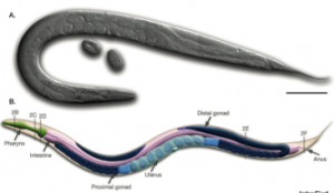
Image 1: Caenorhabditis elegans
On the very first day I learned so many fascinating details about this tiny worm. For example, C. elegans has exactly 959 cells, is normally hermaphroditic, and can take on a Dauer developmental path if conditions are unfavorable. Also, the very first cell division of its embryo is asymmetric (one cell leads to the head and the other to the tail).
Dr. Maddox and Katie are looking specifically at the control mechanisms of the contractile ring during cytokinesis. The contractile ring constricts to pinch a dividing cell in half forming 2 new cells. In the germline of many organisms, the contractile rings halt such that there are cytoplasmic bridges connecting the inner contents of many cells. C. elegans is a great model for studying closing rings and stable rings since the whole organism is transparent and the cells are relatively large.
Questions we pondered:
- How does the cell control the contractile ring?
- Which proteins are involved?
- Can we control these proteins?
- Could manipulating the last step of mitosis be applied to help humans?
Katie showed me how to identify worms in various life stages, pick worms under the microscope, dissect worms to extract embryos, “knock down” targeted proteins to learn function, tag proteins with fluorescent markers, prepare slides for microscopy, and quantitatively analyze resulting images.
Check out some of these pictures from my experience:
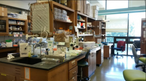
Photo 1: The A. Maddox Lab
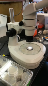
Photo 2: Microscope for picking from plates of C. elegans
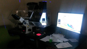
Photo 3: Microscopy in the dark room with a widefield microscope
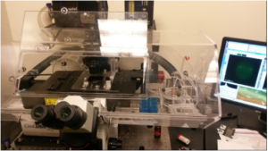
Photo 4: Now with the lights on
Video 1: Click this image for a grayscale video of the first cell division in C. elegans. DNA is tagged in green
Video 2: Click this image for a video showing polarity establishment and cytokinesis. DNA is tagged in red. Polarity proteins, which establish the anterior and posterior ends, are tagged in red and green. Notice the asymmetric cell division.
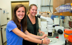
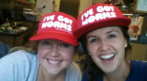
Photo 6: Katie and I doing SCIENCE!
In the final week of my internship, I got to visit various UNC graduate students and postdocs researching other model organisms. I observed and/or learned about yeast, tardigrades, cell cultures, fruit flies, and mice. See the pictures below.
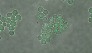
Photo 7: Yeast Budding. An excellent example of asexual reproduction to share with students.
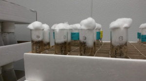
Photo 8: Vials of Drosophila (fruit flies). I learned how to tell the difference between male and female flies, and observed prominent phenotypes like white eyes and curly wings. The kids will love this!
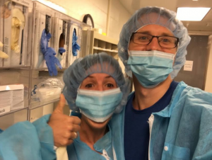
Photo 9: Dressing out with Dr. Andrew Satterlee before a visit to the mouse house. I saw Andrew conduct brain tumor imaging and brain surgery!
Needless to say, science is cool!
Thank you so much to Amy Maddox, Katie Rehain Bell, Kenan staff, and UNC scientists for making my internship experience uniquely wonderful. I’m so looking forward to taking it back to my students!