A-ha Moments

photoDiscover, explore, and wonder! That is my A-ha take away from my Kenan Fellows Museum Adventure this summer.    I had a grand adventure of exploring the outside world, bringing some of it back to the lab to explore some more, and wonder why things are the way they are.

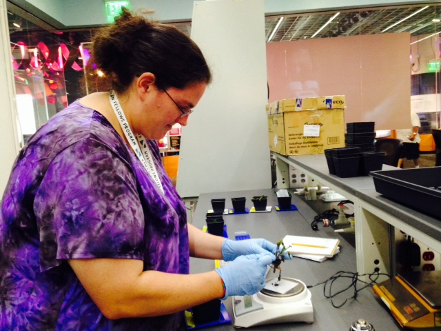
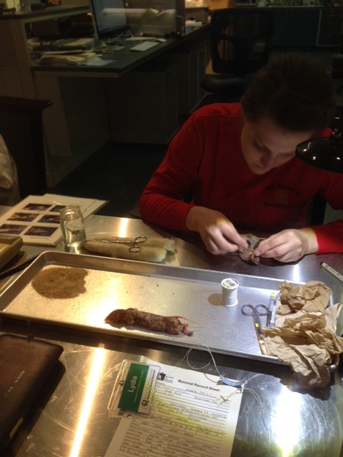
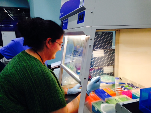
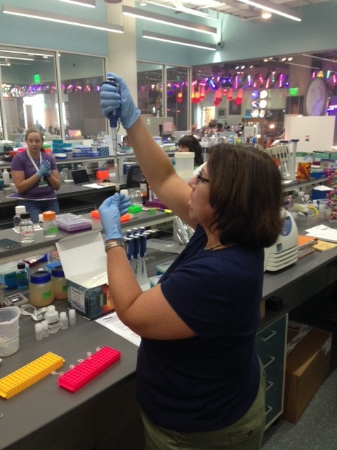
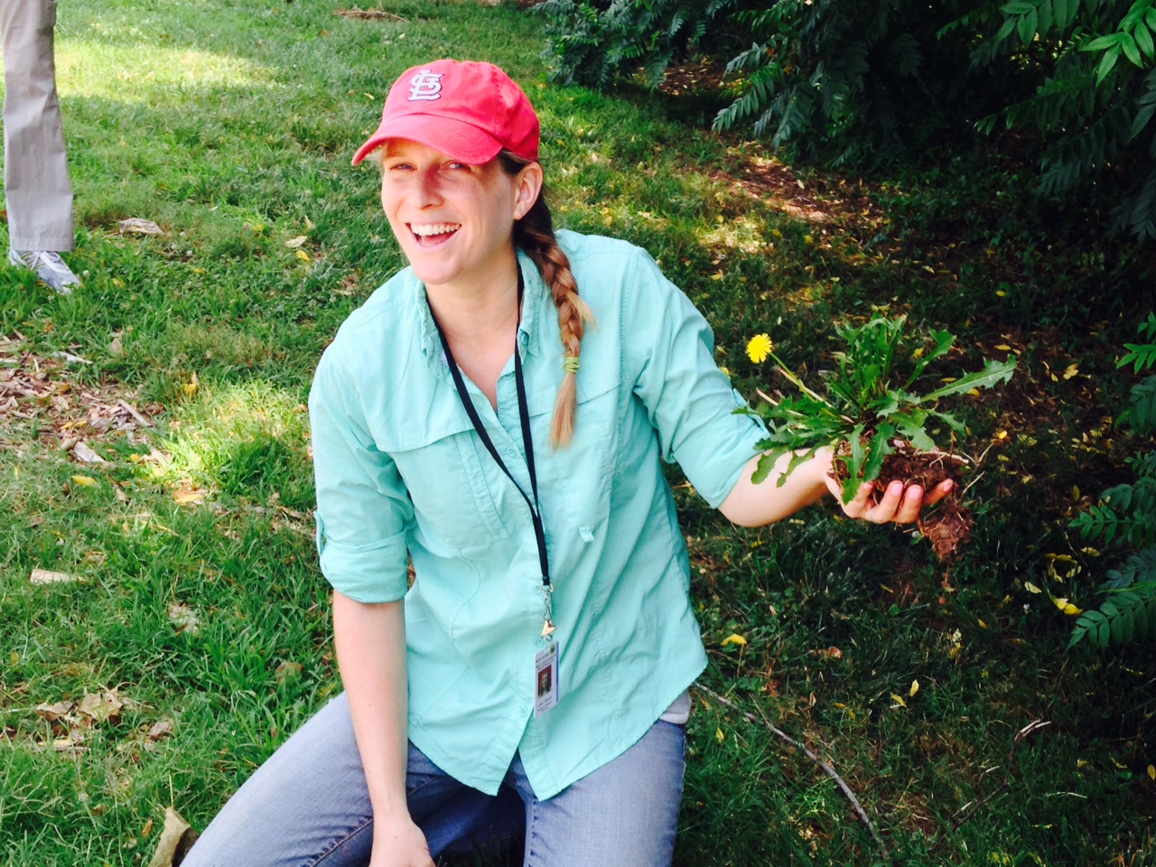
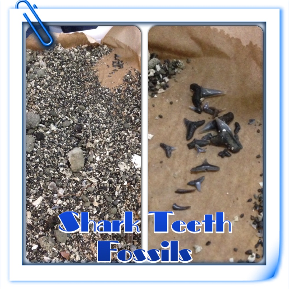
The picture above, of me searching through debris to try and find fossil sharks teeth was me in the middle of one of my A-ha moments.  I realized how much fun I was having searching through to find as many of the teeth as possible, and it hit me that I need to make sure my students experience this sense of excitement in my classroom as often as possible.

Also, going out to Prairie Ridge was a great A-ha moment.  I realized that my students need to go out and be a part of nature. They need to see science and the natural world happening around them. Even, if its something as simple as looking at bugs and watching birds.  Kids need to connect to the outside world. These students of mine are digital natives, and many of them hardly go outside to enjoy just being outside.  But, as I was sitting at Prairie Ridge and using my phone to take pictures, post to inaturalist, and using the Merlin Bird app it hit me, that this is the way to have my students by in to hanging out with nature.  I even went home with my own kids and made sure to enjoy the natural world around them.  We went and explored different geese, fish, dragonflies, and other bugs at a local park.  It was a great time, and I will be sure to give my students some outside time this year.

photo 3          photo 1

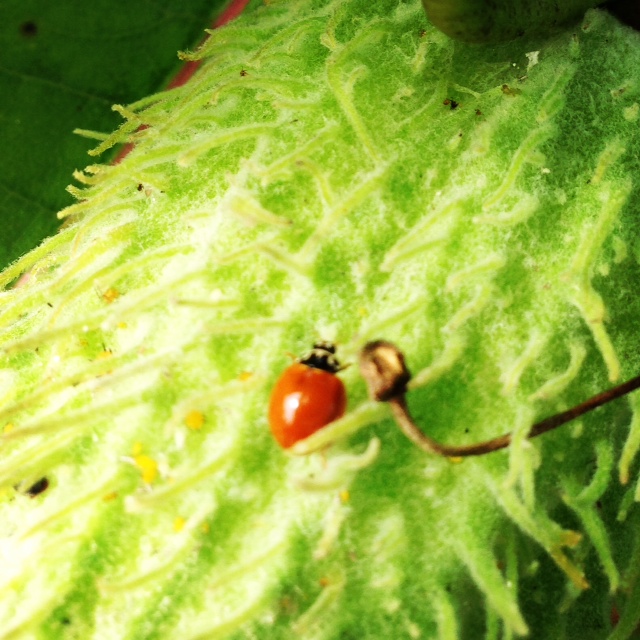
Two of the different Species of Lady Bugs that I hunted down at Prairie Ridge. The Polished Ladybeetle and the Asian Multicolored Ladybeetle.

photo 2

My kids soaking in the wonder of nature.

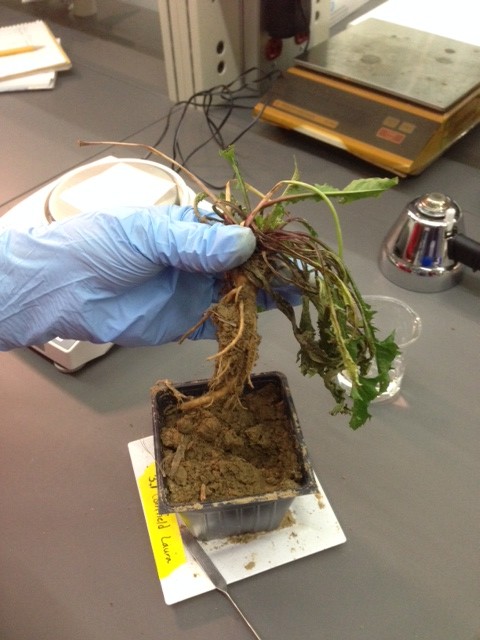
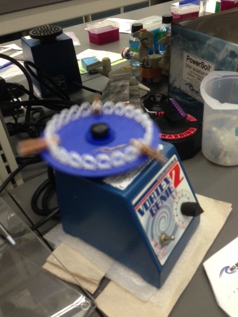
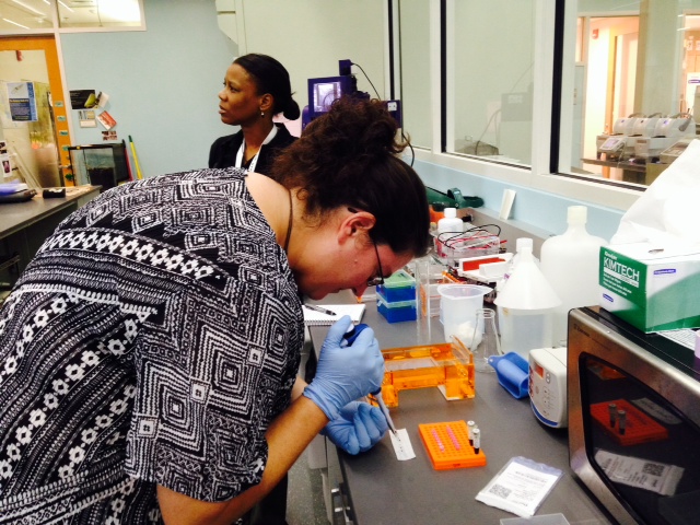
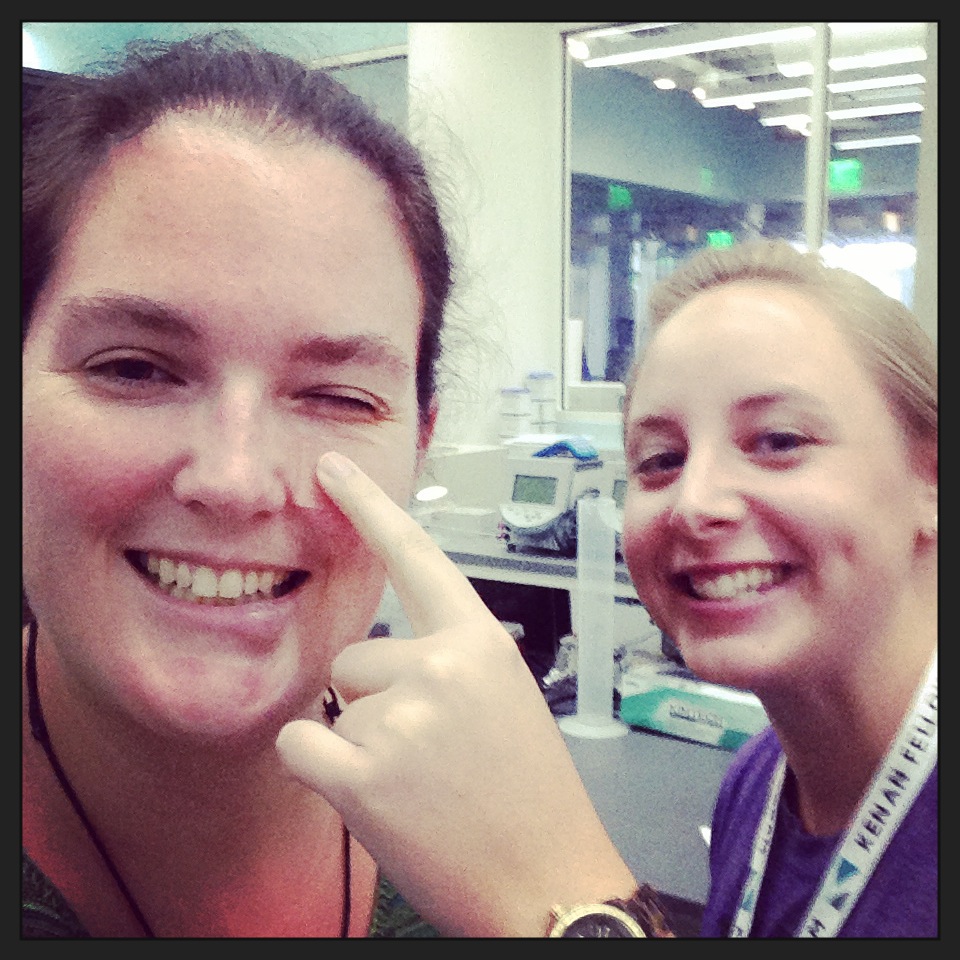
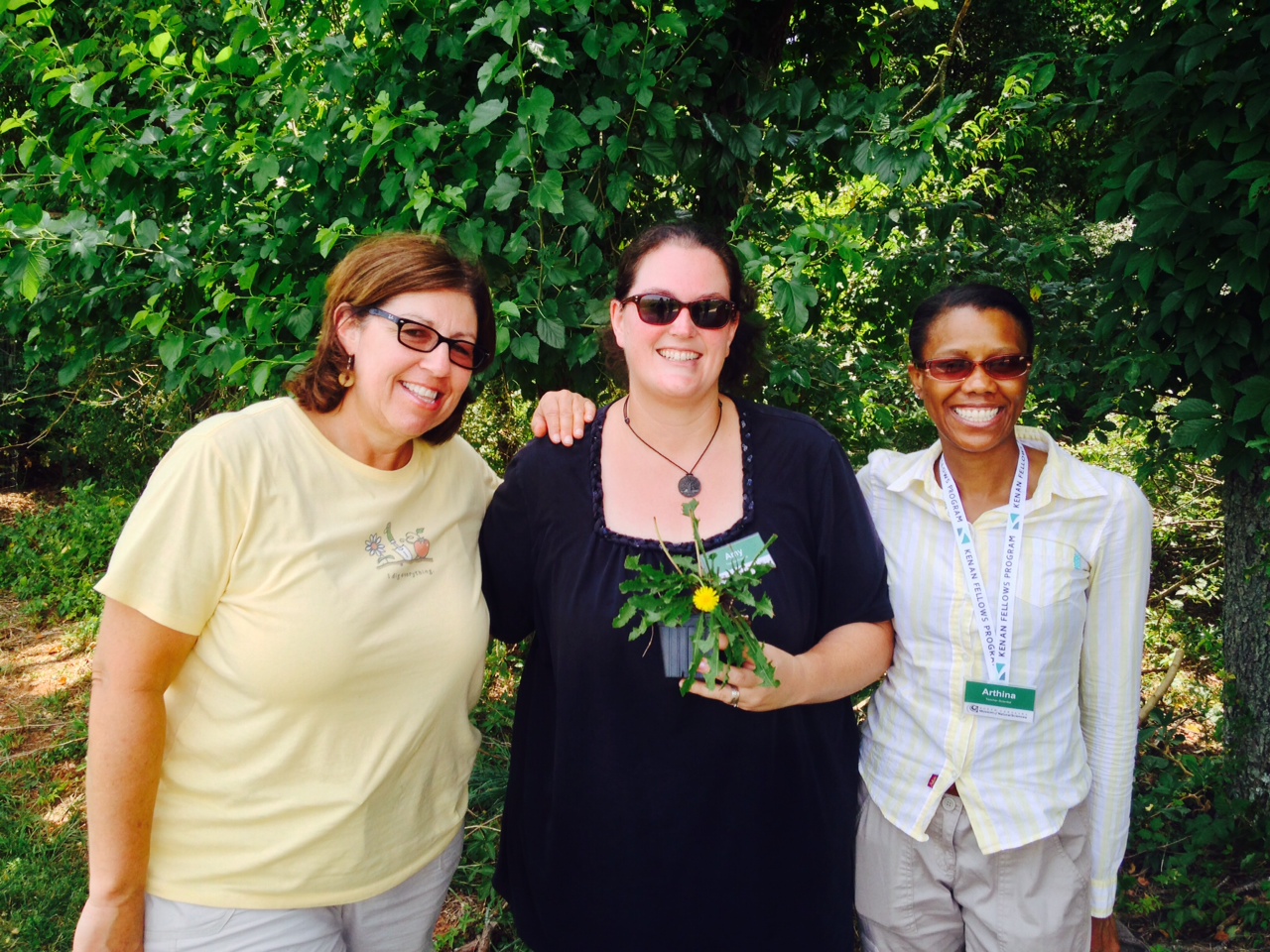
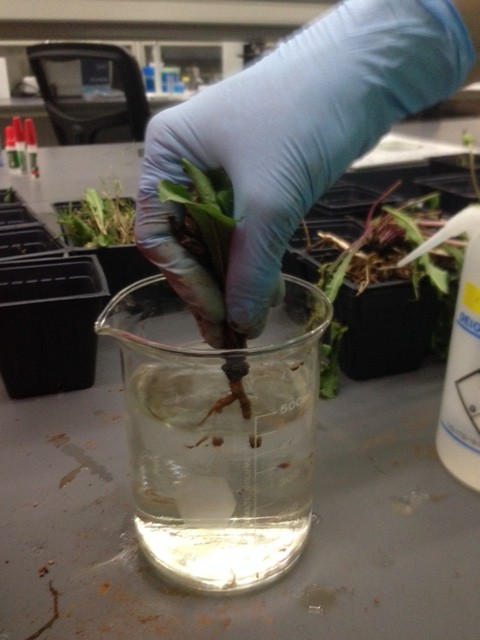
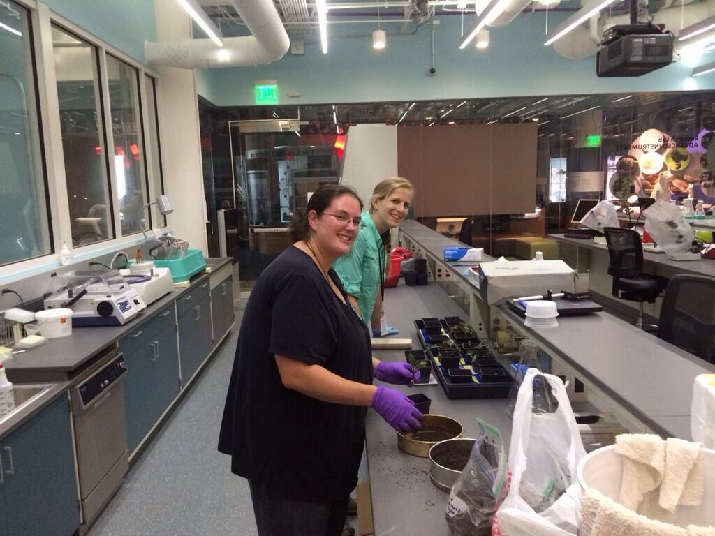
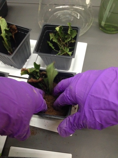
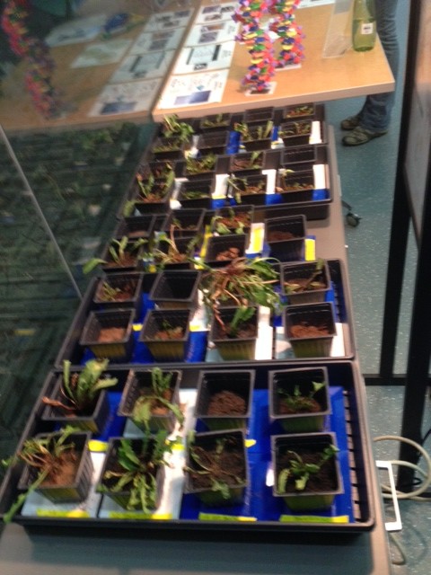
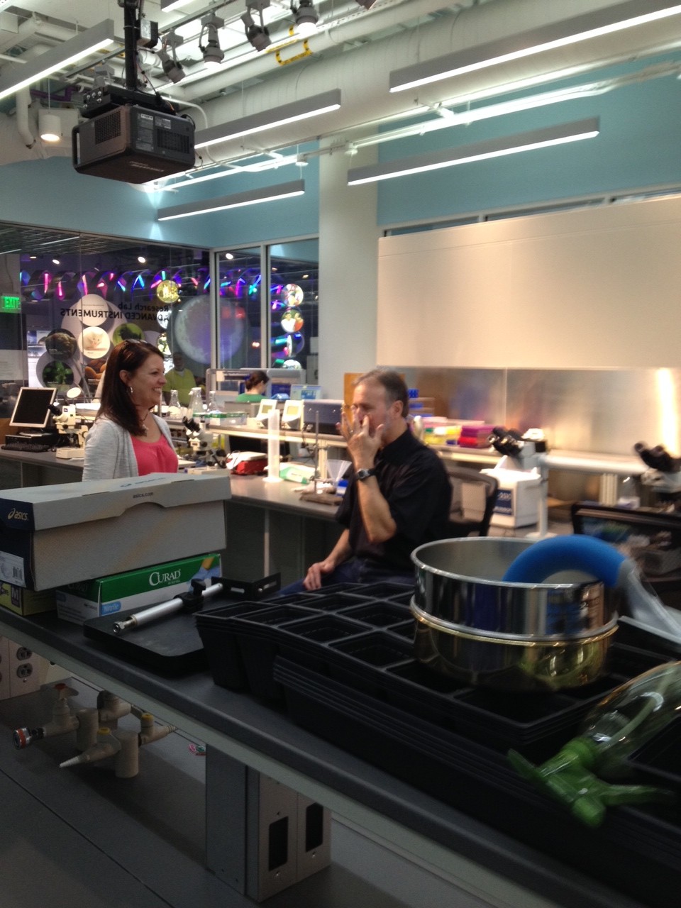
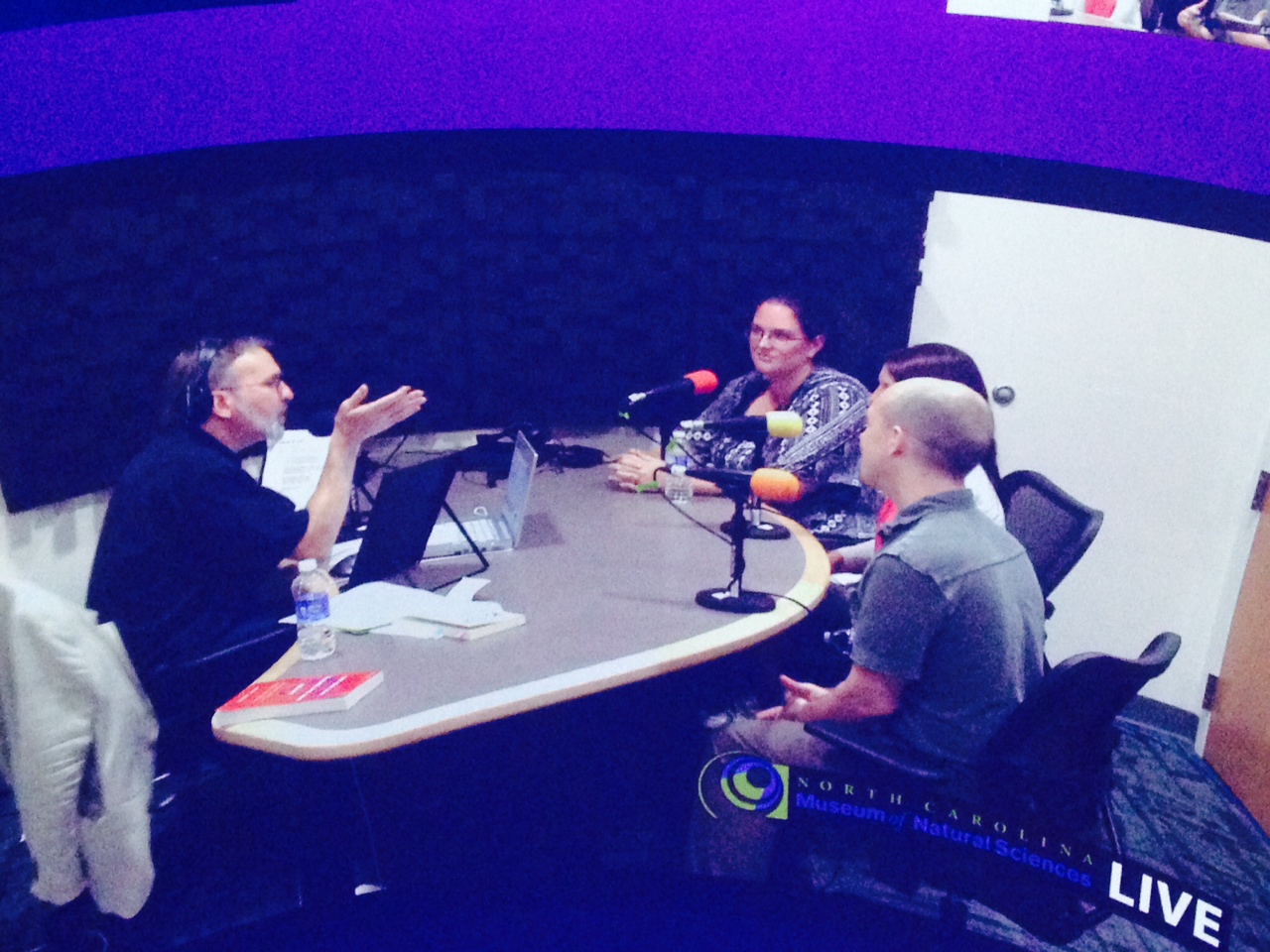
I also had an awesome A-ha moment when I realized how well I worked with my “Team Dirt.” Being able to work in a collaborative group strengthened my ideas, my ability to feel like I could ask questions, and our final work products always seemed to be so much better than if I had done the work all on my own.  This had me realize that I need to have my students collaborate together in my classroom as often as possible. Not just when completing labs or activities, but possibly just every day to just check in with one another about what is going on in the day. It has me trying to brainstorm how I could make this happen.

photo 4

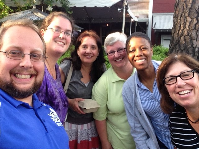
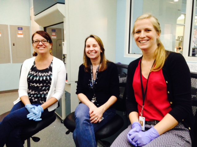
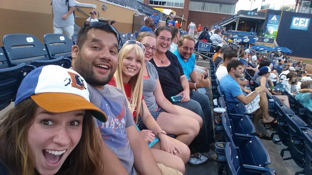
Some of my awesome collaborative teachers and me!

Over all this experience with Kenan Fellows and Students Discover has rejuvenated and revitalized my goals as a teacher. It has made me want to strive to go above and beyond what I’ve already been doing, and truly give my students the best experience in, and out of, the classroom they can get.

Challenges and Success of Curriculum

photo (23)

Building curriculum is often a difficult task, especially when you are trying to make sure it engages students and is completely aligned to the standards.   However, as I have learned through my Students Discover experience, teamwork is an awesome power!!!  At first we struggled with trying to build a lesson that incorporated 6th grade science, 7th grade math, and 8th grade science.  We didn’t know each others curriculum and had a hard time trying to figure how to fit all the pieces together.  So, we had a great idea to put all of our goals together in one document.  That helped us build a bigger and better over all lesson plan that all 3 of could use.  Then, as we continued brain storming, we feel like we have a created an awesome PBL lesson.  It is interdisciplinary and vertically aligned. Having 3 brains working together has been awesome!

The Real Deal!

photo 2 (11)

This week in the lab we are collecting our dandelion project samples that will be sent off to NC State to be run through a MiSeq machine to sequence DNA!  The last two weeks we have been practicing our skills and getting ready for this moment!

We started off the week working with our 39 soil samples.  We have planted our dandelions in triplicate, which means that we have planted 3 dandelions in 3 separate planters for each of the 3 soil samples. We have also put soil into a planter without adding a dandelion for a control.  Each of the three teachers brought in 3 soil samples, so that leads to having 12 samples for each teacher.  Then we collected soil  from Prairie Ridge and kept 3 samples from there.

In order to collect the bacteria and fungus DNA we pulled up soil samples .1 and .2 and scrapped .25g of the soil that was attached to the root of the dandelion. Then on soil sample .3 we did not disturb the dandelion as much, because we just got close to the root without pulling the plant out of the soil.  This trial is called method validation to see if we can get accurate data without uprooting the dandelion.  In sample .4 the soil was just collected, because no dandelions were present.  Each of the dandelions were then replanted in the soil and put back under the grow light and watered to continue growing for the next 3 weeks in the lab.

photo 4 (7)

Me – scrapping soil off of the dandelion tap root.

photo 1 (10)

Show an awesome dandelion taproot and rhizosphere!

Next we began to extract the DNA from all of our samples. We completed 2/3rds of the extractions on Monday and then finished up on Tuesday.  After we completed the extraction we got to use the nanodropper to see if we successfully extracted the DNA before we put it into the PCR machine to duplicate the DNA. We were all a bit nervous, because this was the real experiment and not just practice any more. We were all very excited when the nanodrop showed all of our samples had a very nice concentration of DNA, so we set up the samples and ran the samples through the PCR machine.

The cool part of setting up these samples was that we were using a new “Master Mix” that cut out some of the pipetting steps. We also were adding in adaptive primers to allow our DNA samples to be sequenced in a high tech DNA sequencing machine called a MiSeq.  This machine is housed at NC State and will allow the genomics lab to create 15 million copies of DNA in one run! This technology is amazing, and only costs about a thousand dollars.  This is comparatively inexpensive when compared to just a few years ago where labs would have to spend tens of thousands of dollars or hundreds of thousands of dollars to duplicate DNA in much smaller quantities.  Team Dirt is on the cutting edge!!!

photo 3 (7)Dr. Urban, Dr. Horvath, and Dr. Stevens as they help us through our DNA extraction.

photo 3 (9)Using the Vortex machine to begin extraction of DNA.

photo 1 (12)Our 39 samples of extracted DNA.

We were all very excited to see our electrophoresis gel after such a success on nandrop, but alas……our gel showed that the DNA was not duplicated correctly. All 4 of us on Team Dirt were very sad, but we continued on.  We knew that failure is what science is all about! We learn from our failures, and that is what we did.

Dr. Stevens ran a short series of trouble shooting experiments with our extracted DNA.  She picked 5 random samples out of our 39 and then retested them 4 ways.  The first two trials used the “Master Mix”. Trial 1 used the MiSeq adapter primers, Trial 2 used the non-adapter primers.  Trials 3 and 4 did not use the “Master Mix” and instead each individual ingredient, including the Taq was added by hand.  Then Trial 3 used non-adapter primers and Trial 4 used adapter primers.  After these trouble shooting experiments were run, and we got to look at the gel, we saw that the “Master Mix” did not work very well with our soil DNA, but that our hand made mix was giving us very good readings. This made all of us on Team Dirt very very happy! We now know that we can take all 39 samples of soil DNA and remix them and prepare them for PCR machine and being shipped off to NC State for the the MiSeq machine!!!!

photo 2 (9)

Me working on setting up PCR.

photo 1 (11)Our Trouble Shooting Electrophoresis Gel

Today our team also plated soil extracted from all .3 plants on various agar. We have our experimental soil extracted agar plates that we created last week, as well as nutrient agar and bright pink fungal plates! We are going to grow the bacteria and fungus on these plates and take pictures of the growth each day, so that students can visually see species diversity and how it can grow and change over time.

This week Team Dirt has also been working on building our curriculum from the ground up. It has been challenging, but we are determined to build a fantastic PBL lesson that will be and excellent Citizen Science project.

Random Shots from the Week

photo 3 (8)Our Sign in the Lab Window!

photo 5 (8) photo 4 (8)A storm approaching and reaching my 17th floor room.

photo 2 (10)The Biodiversity lab preserved multiple animals from collections in the lab this week. This is a chipmunk above.

Classroom Curriculum Connection

This Kenan Fellowship and Student Discover adventure at the Museum has been a great time to rejuvenate my teaching soul.  I’m getting excited about building projects for my students to complete as scientists. Team Dirt and I have been talking curriculum all week. Yesterday we hashed out quite a few ideas and have nailed quite a few of them into place,  at least with one nail, hammered in about half way……

So far we known that students at Mills Park Middle School in Wake County will be on an Edmodo with Burgaw Middle School students in Pender County.  In Edmodo the students will be able to discuss and share their awesome adventures into Citizen Science. They will also have assignments that must be completed and turned in through Edmodo.  This part of the lesson will teach students about their global world. They will be able to connect with other students in their state, and they will be able to talk about their science experiments as scientists.

Another way that students will see how they are connecting to their community and global world is by getting to meet the scientists that work in the lab. Next year in our classes the students will get to meet Dr. Julia Stevens when she comes to visit our schools, but they will also get to use a distance learning tool to video conference with Dr. Stevens and Dr. Urban in the museum lab. Students will get to talk and interact with the scientists and see their own personal results being shown to them from within the lab.

The next part that we have decided upon, is to try to make our project a Project Based Learning experience that can be used as an interdisciplinary unit if teachers of all subjects will buy in to the lesson and help.  Students will be doing science experiments in science class.  This experiment will have the students bringing soil from their communities into the classroom to find out what bacteria and fungi are present, and which ones dandelions will gain in their own “microbiome.” Students will then go around the school and collect dandelions to sterilize their roots and replant into the student’s soil.  Then the students will grow and observe the dandelions until we collect the roots and the soil to be sent back to the museum lab for testing.

As the students are growing the dandelions we will be able to discuss and learn about photosynthesis, the nitrogen cycle, symbiosis, harmful and helpful bacteria, food webs, ecosystems, populations, species, and taxonomy.  All of these things will be explored in our research project, and all of them tie in with the current 8th grade NC Science Essential Standards.  We will also be able to tie in soil, photosynthesis, and biomes which are  in the 6th grade NC Science Essential standards  Also, there is a lot of ratios, percents, graphing, scatter plots, and quadratics which tie in to 7th Grade and Math 1 standards.

DNA Electrophoresis, Beautiful Bacteria, Butterflies, and Symbiots

photo 1 (8)

The last two days in the lab have been spent on learning how to make a gel for electrophoresis.  Electrophoresis allows us to put our DNA into a gel, run electrcity through a solution, and see if we replicated the correct DNA strands for sequencing.  Our first try at electrophoresis  failed, because we did not put in a key ingredient to the solution we put into the gel, but when we tried it the second time, it worked beautifully!

photo 2 (6)

Me mixing DNA Extraction mixture  and Electrophoresis Dye

photo 5 (6)

Close up of the mixing

 photo 1 (9)

Laura putting the Mixture into the Gel

photo 2 (8)

Our successfully filled Electrophoresis Gel!

photo 4 (6)

Electrophoresis Machine Running!

photo 3 (5)

Our successful gel read!

Yesterday we looked at our agar plates full of beautiful bacteria. It was fun getting to look at all of the different species of bacteria growing on the plates. It was amazing see all of the different types in such a small area. We believe this will be a great way to show students about diversity of species.

photo 5 (5)

One of our cool bacteria plates

We also had 2 fun excursions out into the museum. The first excursion was a nice relaxing trip up to the butterfly house. I loved seeing all of the beautiful butterflies flit around my head. Then we got to sit in on a cool Daily Planet Talk by Dr. Julie Urban. She talked about how animals and bacteria life in symbiosis with one another. It was fascinating to learn how planthopper insects make cells in their bodies to stick bacteria into, and then they become permanent appendages. Crazy! She also talked about the Hawaiian Bobtailed Squid and how it uses bioluminescent bacteria inside of their eyes as counter illumination to defend against predators! So Cool!

photo 2 (7)

Longwing Butterfly

photo 3 (6)

 Postman Butterfly

photo 4 (5)

Julie giving her symbiosis Daily Planet Talk

week-in-wildlife-The-Hawa-001 squid2

The  Hawaiian Bobtailed Squid

Extracting DNA, Duplicating DNA, and Face Mites

photo 5 (3)

Today we started the day learning the specifics on how to use the PCR (polymerase chain reaction) machine to duplicate DNA.  Then we got to extract the DNA from soil we collected around downtown Raleigh. All 3 of us on Team Dirt had an extremely successful first try at DNA extraction! We were shown how to use a cool machine called the nanodrop to test the concentration of extracted nucleic acids, and we all got good levels of concentration! Then we went on to prep the PCR  mixture and put it in the PCR machine before we left for the night.

photo 5 (4)

Laura working on her DNA extraction.

photo 3 (4)

Dr. Stevens drops our extracted DNA onto the NanoDrop machine.

photo 2 (5)

Sterilizing our equipment with UV rays.

photo 1 (5)

Using the Crosslink UV sterilizer.

photo 4 (3)

Putting our extracted DNA into the PCR mixture.

photo 3 (3)

Into the PCR Machine we go!

As I was about to leave the museum for the night I noticed that Team Face Mite was still working in their lab, so I went in to see what science they were up to.  They were scraping Kenan Fellows for mites, and doing super glue extractions of mites. So I jumped on in and had them check my face for mites, and WOW! I had mites! They found an adult and juvenile together and a few other mites just in a small 1 inch square sample on my face. It was amazing to see these little creatures that live on my face under the microscope!

photo 2 (4)

Meribeth on Team Face Mites removing them from my face!

photo

One of my face mites!

Flower Picking, Anesthetized Fish, a Display in a Museum, and the Durham Bulls!!!

 photo (21)

WOW! What a day! We started off the day hunting for dandelions out at Prairie Ridge.  It took us a while to find them, but eventually we did!  Team Dirt dug up 30 dandelions and brought them back to the museum! After we brought them back to the museum Julia sterilized their roots in a 10% bleach solution and we got to replant the dandelions in our soils that we brought from home! We are all excited to get to see if our dandelions recruit some of the same symbiots to live in their microbiome.

photo (20)

Doctor Julia Stevens so excited to get a blooming dandelion!

photo 1 (2)

Sterilizing roots in 10% bleach solution

photo (22)

Sifting my soil to plant my dandelions

photo (18)

Planting Dandelions!

In the middle of sterilizing the roots and planting the dandelions we got to go visit the museum’s vet lab, where we got to see them look for mycobacterium on a Black Banded Sunfish.  It was so neat getting to watch them anesthetize  a fish and take scale, gill, and fin samples.  Then we got to see them look at the samples up on a live microscope screen.  The whole time we got to hear what the  Vets were saying and ask them questions through microphones on the inside and out.  So cool!

photo 5 (2)

After our visit with the vet we went back to the lab to continue setting up our experiment and a display to put in the front windows of the Genomics Lab.  By the end of the day the experiment was all set up and Team Dirt has an official display in the museum!!!

photo (17)

Team Dirt’s Display

photo 1 (3)

Our Test Subjects!

photo 2 (3)

Our Display!

After I left the museum for the evening I met up with some other Kenan Fellows to go to a Durham Bulls game! A home run was hit, and we got to see the famous Durham bull’s eyes light up and smoke come out his nostrils! And to top it all off the Bulls won! Yay!

photo 4 (2)

photo 3 (2)

Tech Challenges

In education there are 3 main issues with using technology effectively: time, money, and understanding.

Time with the technology is usually very limited for our students.  At our school we are allowed to take our students to the computer lab once a week.  It works to give them  a chance to be exposed to the technology and we can do short technology lessons. However, it does not lend itself to doing any sort of long term project based around technology.  Another problem was when our school first got in a cart of I-pads, we had to figure out how to share, you were lucky to be able to get to use them for a whole day.

Technology also costs the school systems a lot of money.  Many of the desk top computers that we have in our school are between 10 – 5 years old, because it is very costly to replace them.  Also many of my students do not have money to access technology at home either.  I am at a Tittle I school with over 80% of the population having free and reduced lunch, so technology is not always a priority at home.  I have been blessed the last year, because our school has been using our money to buy new Tech.  We have chosen to try and get technology above and beyond anything else.  In fact, our whole count is going 1 to 1 with devices for our students on the middle school level next year.  I am very excited to begin the school year knowing that our students will have access to a chromebook all day long.  However, I know that this seems like a dream to many of the counties around us.  I hope that money can be found for all school systems to be able to venture into the 1 to 1 classrooms.  Another part of technology that we do not have money for is the higher end science technology.  In my project we are using Autoclave machines to sterilize materials, PCR machines to duplicate DNA,  incubators to grow cultures, microscopes to view microbes, DNA sequencing machines, and even basic technology like pipettes.  All of these items are costly, some beyond even imaging a school could purchase.  So, we as teachers are going to have to come up with creative ways to adapt experiments or how to partner up with our local universities and research labs to help our our young middle school scientists.

Understanding how to use the technology does seem to be a barrier for both teachers and students.  When we have lots of different technology in a school its hard to teach everyone how to use the different brands and types. We have Desktop PCs, I-pads, I-pod touches, Chrombooks, Netbooks, white boards, and mimios all in my school.  For some people that are tech savvy its not a problem to understand how to use all of the different types, but some teachers and students get confused by the different interfaces.  So time building lesson plans or time teaching is taken away to learn how to use the technology.  When we spend half the class trying to get the students to remember how to log on to a new device, or get it connected to the wireless routers, we lose teaching time.  This is why, I am super excited to know that all of our students will be on the same brand and type of device next year. Time will be lost at the beginning of the year teaching the students how to use the device, but then as the weeks go by, it will be as easy for them to use as pencil and paper.

Genomics Lab and NPR

20140702_120526-1

Dr. Urban, Dr. Dunn, and Me

This has been a very eventful two days! Yesterday and today we got into our labs to learn how to use all of the different tools, and we even got to use some of them! Team Dirt got to make our own Nutrient Agar Plates, collect soil around downtown Raleigh, add soil to Phosphate buffered solution (PBS), centrifuge the mixture, pipette the solution with bacteria cells on to the agar plates, and incubate the bacteria! We can’t wait to see what is growing on our Agar tomorrow!!! We have also been taught by our Mentors about how we are going to extract DNA, duplicate it, and sequence the DNA with next gen technology.

photo (14)Practicing my pipetting skills in honor of the USA Soccer Team!

photo (15)

Our nutrient agar plates.

photo 2 (2)

Me pipetting out PBS solution and soil bacteria mixture.

20140702_165940

Me, Laura, and Arthina at work in the Genomics Lab!

We have also be able to listen to the Post Docs present their projects to the Museum and all of the Kenan Fellows.  After the post docs presented their materials we were allowed to get a behind the scenes tour of their labs. Yesterday we got to visit the Paleontology Lab, where we saw many dinosaur fossils and shark teeth.  I found it fascinating to see how carefully the fossils were cleaned and exposed. It reminded me how science is a craft of patience.  I also thought it was amazing that a new Dinosaur was discovered by Dr. Gates, and he is getting to name it! I can only dream that Team Dirt discovers new bacteria or fungus. That would be an amazing story to tell my students.

photo (16)

 Dr. Dan Fergus presenting his lesson on face mites.

photo (12)

Carefully separating plaster, rock, and fossil from dinosaur bones!

photo (13)

We got to test out the debris that our students will get to use in the paleontology Student Discover project. 

 Today I was also given the opportunity to be a guest on the NPR show, “The State of Things”, hosted by Frank Stasio. I accompanied Dr. Robert Dunn the lead investigator on our grant and Dr. Julie Urban the Assistant Director of the Genomics Lab. This was an amazing experience! I’ve never been on the radio before, and I was glad that I got to represent the 12 Student Discover Kenan Fellows.

photo 4 (1)

Mr. Frank Stasio removing his mite from his face before the radio broadcast.

photo 3 (1)

Mr. Stasio, Dr. Dunn, Dr. Urban, and Me.

photo 1 (1)

Me  being projected up on the big Globe Theater  Screen inside the Museum!

What a great 2 days!